
Fig 1. Leica DVM6 microscope
Fig 1. Leica DVM6 microscope
This microscope has a built-in 10MP camera (3664 x 2748 pixels) and has three choices of objective lens module. The module used for this analysis was for 'medium magnification' (42x to 675x), Leica PlanAPO FOV 12.55 (10 450 705).
As per the Focus Analyser procedure, the test target for these analyses was a carbon-soot-coated straight-edge razor blade inclined (with respect to the optical axis) by 6.5° and tilted (with respect to the camera frame) by ~9°, elevated about 5mm above an out-of-focus white paper background. The target was illuminated obliquely on two sides by fiber optic illuminators.
I had only two hours with the microscope to become familiar with it and obtain images, without a manual. Alas, there are two problems with the images obtained: 1) the images are 2MP (1599 x 1199 pixels) due to mistakenly capturing the preview image instead of the full 10MP image; 2) the images were sharpened by the Leica LAS software, as evidenced by the overshoots bordering the edge on the 'edge profile charts', despite sharpening being set to 'off' (perhaps the setting only affects the capture image, not the preview image). There didn't seem to be way offered by the Leica LAS X software to obtain raw image files.
At 2MP, the resolution of the images may not be sufficient to capture the detail provided by the microscope. True resolution is further obscured by sharpening, which makes edges appear sharper. In sum, probably the results below provide only 'ballpark' values on the microscope's resolution. However, the information about longitudinal chromatic aberration is probably reliable.
The image files are assumed to be gamma-encoded. 'Zoom' figures are as displayed by the microscope interface.
Below are Focus Analyser reports for selected zoom settings. The field of view (FOV) for each analysis was determined by taking a photo of a scale positioned perpendicular to the optical axis at each zoom level. The scale in the image was approximately 1.6% longer than the scale generated by the microscope LAS X software; the real-world scale was used below.
Ideally, the microscope + camera adapter would focus all three colours identically; the curves would overlap. Generally the curves do overlap, as expected with an apochromatic lens, until the highest zoom, where the blue channel diverges from the green and red (behaving like an achromatic lens).
The asymmetry of the 86x and 200x results suggest the point of best focus wasn't at the center of the inclined straight edge (rather, it was slightly to the left).
Figure 3 below charts resolution (as obtained from the edge spread analyses above) vs field of view. Recall that these values have been improved by sharpening, making the resolutions better (lower) than true. The red line indicates the Nyquist resolution limit associated with the camera's resolution. Resolution better than this might be measurable via subsampling, but more likely the results have been affected by sharpening interfering with the edge analysis.

Fig 3. Resolution (μm) vs field of view (mm). Red line is the Nyquist limit. Sharpening has caused measurements to be better than true.
Leica says the expected resolution is 11.1 μm and 0.93 μm (corresponding to line pair / mm values of 90 and 1073) at 86x and 1422x respectively, which are close to but slightly better than the results above. Images of random patterns (scratched metal) yielded resolutions via the FFT method somewhat lower (26 and 1.7 μm), perhaps because images of random patterns are not easily sharpened by software. This could be clarified by obtaining images with the camera's 10MP capability.
The published resolutions provide some reassurance that the 2MP images were sufficient to capture meaningful data, especially away from the point of sharpest focus; sharpening during post-processing was likely more of an issue.
Figure 4 is a crop viewed 1:1 pixels of a portion of a 2MP photo of an integrated circuit, lit by co-axial lighting, at 1422x. The teeth in the comb-like features are spaced by approximately 11 microns. This is the highest quality image I've seen of this object (compare, for example, with results from a modest stereomicroscope or a good macro lens).

Fig 4. Cropped view at 1:1 pixels of a portion of a 2MP photo at 1422x of an integrated circuit, lit co-axially.
Figure 5 is another view of the wafer, at about 200x (click for full image including a scale bar). The resolution is probably limited by the image size rather than the microscope optics.

Fig 5. Cropped view of a portion of a 2MP photo at approximately 200x of an integrated circuit, lit co-axially.
Figure 6 is a composite image of aeolian sand grains, created by focus stacking (without which only a shallow layer would be in focus). Click for full image including a scale bar. In this case the image produced by stacking is the full 10MP (and likely contains some 'empty magnification').
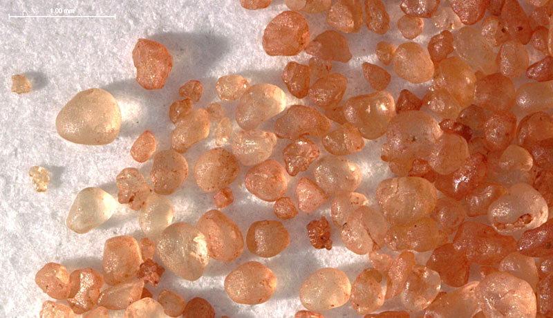
Fig 6. Cropped view of a portion of a 10MP photo at approximately 100x of aeolian sand grains, lit obliquely.
The Leica microscope and software excel at making such imaging easy and quick, requiring just setting the focus range and then a mouse click to initiate the process. What might take hours manually takes only seconds.